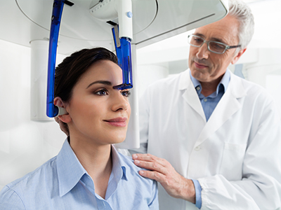
At Restoration Dental, we incorporate advanced imaging to make diagnosis and treatment planning more precise, predictable, and comfortable. Cone-beam computed tomography (CBCT) gives our clinicians high-resolution, three-dimensional views of the teeth, jaws, and surrounding structures that traditional X-rays cannot provide. When used thoughtfully, CBCT helps uncover anatomy and relationships that are critical to safe, effective dental care.
CBCT is a focused 3D imaging technology designed specifically for dental and maxillofacial applications. Unlike conventional medical CT scanners, dental CBCT machines capture detailed volumetric data with a single short scan and specialized software that reconstructs the images for clinical review. The result is a clear, spatial view of bone, tooth roots, nerve pathways, and sinus anatomy that supports more informed clinical decisions.
We prioritize patient safety and comfort during imaging. Modern CBCT units deliver targeted scans that limit radiation exposure to the area of interest while producing diagnostic-quality images. Our team follows established protocols to ensure each scan is necessary, properly positioned, and interpreted by experienced clinicians, so you get the maximum benefit with the least exposure.
Three-dimensional imaging expands the clinician’s ability to detect conditions that can be missed or obscured on two-dimensional X-rays. Small fractures, complex root anatomy, impacted teeth, and the precise spatial relationships of lesions become visible in CBCT volumes. These insights can change a diagnosis or reveal considerations that alter the treatment approach.
For periodontal and endodontic evaluations, CBCT helps clinicians see the exact extent of bone loss or infection pathways. For oral surgery, the scan shows where important nerves and sinus spaces lie in relation to planned procedures. This ability to visualize structures in three dimensions reduces uncertainty and supports safer, more conservative care plans.
CBCT also aids in the detection and assessment of developmental and pathological findings, such as cysts, unusual tooth positions, or abnormal bone growth. Early identification of these issues allows for timely intervention and coordination with specialists when necessary.
Implant placement is one of the most common applications of CBCT in dentistry. The technology lets clinicians measure bone volume and density in three dimensions, identify vital structures like the mandibular nerve and maxillary sinus, and plan the ideal implant size and position before any surgical work begins. That level of planning reduces surprises and helps achieve consistent functional and aesthetic outcomes.
When combined with digital prosthetic planning tools, CBCT data can be used to design surgical guides that translate the virtual plan to accurate placement in the mouth. This integration supports minimally invasive approaches, predictable healing, and a higher likelihood of long-term implant success.
CBCT is also valuable for evaluating potential grafting needs and assessing the quality of existing bone. Knowing what to expect before entering the surgical field streamlines procedures and helps clinicians communicate realistic steps and timelines to patients.
Safety is a primary benefit of modern imaging. CBCT reveals critical anatomical landmarks and variations that, if unrecognized, could increase the risk of complications. Identifying the position of nerves, the depth of sinuses, and the location of adjacent tooth roots allows for well-informed surgical decisions and reduces the likelihood of intraoperative surprises.
Beyond landmark identification, CBCT can be used to map trajectories for implant drills and to verify the relationship of pathology to surrounding tissues. For patients with prior surgeries, retained root fragments, or complex anatomy, a scan provides a reliable roadmap that enhances clinical judgment and procedural confidence.
Because radiation dose and image field size are controllable, clinicians can tailor scans to the clinical question—scanning a single tooth region when appropriate rather than capturing a broader area. This targeted approach balances diagnostic needs with the principle of keeping exposure as low as reasonably achievable.
CBCT images are valuable for coordinated care among general dentists, oral surgeons, periodontists, and endodontists. When multiple providers review the same 3D dataset, they can discuss findings with a shared visual reference, which streamlines referrals and collaborative treatment planning.
For orthodontic cases and airway assessments, CBCT provides information about jaw relationships, airway volume, and tooth positions that supports comprehensive planning. Similarly, in restorative dentistry, three-dimensional data helps ensure that crowns, bridges, and full-arch solutions are designed to fit the available anatomy and occlusion.
Because the scan can be archived and re-evaluated, CBCT also creates a permanent record that clinicians can reference throughout a treatment sequence. This continuity supports informed decision-making at each stage of care.
A CBCT appointment is typically brief and noninvasive. Patients are positioned in the unit and instructed to remain still for a single short rotation, often lasting less than a minute. The scanner captures a volume that is then reconstructed into multiplanar views for clinical review. No injections or sedation are required for the imaging itself.
After the scan, the clinical team reviews the images and integrates key findings into the treatment plan. Because CBCT produces multiplanar and 3D renderings, clinicians can measure distances, review cross-sectional slices, and isolate areas of interest with precision. This information informs surgery, restorative design, and any recommended referrals.
We ensure that each scan is interpreted by clinicians trained in CBCT review and that patients receive clear explanations of what the images show and how the findings influence care. When appropriate, clinicians will share annotated images to help patients understand their condition and the rationale for recommended steps.
CBCT is a tool that supports better outcomes when used judiciously. If your provider recommends a scan, it’s because the additional detail will improve the safety, accuracy, or predictability of your treatment.
In summary, cone-beam computed tomography is a powerful diagnostic resource that enhances our ability to plan treatments safely and effectively. The practice uses CBCT selectively to gain critical insight into anatomy, minimize intraoperative risks, and improve restorative and surgical precision. If you would like to learn more about how CBCT may be used in your care, please contact us for more information.


Cone-beam computed tomography, commonly called CBCT, is a focused three-dimensional imaging technique designed for dental and maxillofacial applications. The scanner captures volumetric data in a single short rotation and software reconstructs detailed cross-sectional and 3D views of teeth, bone, nerve pathways and sinuses. These volumetric images reveal spatial relationships and internal anatomy that two-dimensional X-rays cannot show reliably.
Compared with conventional dental X-rays such as bitewings or periapicals, CBCT provides depth and multiplanar detail rather than a flat projection. Unlike full medical CT systems, dental CBCT units are optimized to image the jaws with lower radiation and higher spatial resolution for hard tissues. That combination makes CBCT particularly useful when clinicians need precise information about bone shape, root anatomy or adjacent anatomical structures.
CBCT is recommended when three-dimensional detail is likely to change diagnosis or treatment planning, such as implant planning, surgical extractions of impacted teeth, complex endodontic assessment, and evaluation of cysts or abnormal bone growth. It is also useful for assessing bone volume and the relationship of teeth to nerves and sinuses before invasive procedures. In orthodontics and airway evaluations, CBCT can provide additional information about jaw relationships and airway space that complements clinical examination and 2D records.
At Restoration Dental, clinicians select CBCT selectively rather than routinely, using the scan when the additional information will improve safety, predictability or outcomes. The decision to scan is based on the clinical question, patient history and the potential value of three-dimensional data to guide care. When a scan is recommended, the team will explain the reason and how the images will be used in planning treatment.
CBCT allows clinicians to measure bone height, width and density in three dimensions, identify the exact location of vital structures such as the mandibular nerve and maxillary sinus, and evaluate the quality of available bone. This information supports selection of appropriate implant size, angulation and depth before surgery, reducing intraoperative surprises. When combined with digital restorative planning, CBCT data helps coordinate prosthetic goals with surgical positioning to achieve predictable function and esthetics.
CBCT datasets can also be used to design and fabricate surgical guides that translate the virtual plan into precise clinical placement. Guided surgery based on CBCT reduces guesswork, enables minimally invasive approaches, and improves reproducibility of implant position. In addition, scans help clinicians assess whether bone grafting or additional procedures are required and provide a roadmap for those interventions.
Modern dental CBCT units are designed to deliver targeted scans that limit radiation to the area of interest, and clinicians follow the ALARA principle—keeping exposure as low as reasonably achievable. Field of view, resolution and scan time are adjusted to suit the clinical question so that only the necessary anatomy is imaged. Compared with medical CT, dental CBCT typically uses lower radiation for comparable anatomic regions, but any imaging decision balances diagnostic benefit against exposure.
Prior to scanning, the clinical team evaluates whether CBCT is essential for diagnosis or planning and discusses alternatives when appropriate. Patients should always inform the team if they are pregnant or believe they may be pregnant so the provider can adjust the approach. Protective measures such as thyroid collars, limiting repeat scans, and careful positioning further reduce unnecessary exposure.
A CBCT appointment is usually brief and noninvasive, often lasting only a few minutes from positioning to image acquisition. The patient is positioned in the unit—either sitting, standing or stabilized in a supine position depending on the device—and asked to remain still while the scanner rotates to capture the volume. There are no injections or sedation required for the scan itself, and most patients experience no discomfort beyond maintaining stillness for the short scan.
After acquisition, the scan is reconstructed into multiplanar views and 3D renderings for clinical review, which may take a short additional processing interval. The provider will review the images, annotate findings when helpful, and incorporate them into the treatment discussion. Patients are encouraged to ask questions about what the images show and how the results influence recommended care.
CBCT images are interpreted by trained clinicians with experience in three-dimensional dental imaging; in complex or ambiguous cases, images may be reviewed by an oral and maxillofacial radiologist for additional expertise. Interpretation involves reviewing axial, sagittal and coronal slices as well as 3D renderings to assess anatomy, pathology and relationships relevant to the clinical question. Clinicians use measurement tools, cross-sectional views and annotations to document findings and support precise treatment planning.
Clinicians communicate key findings to patients in clear, nontechnical language and often show annotated images to help explain the anatomy and rationale for proposed treatment. When referrals or interdisciplinary coordination are needed, the CBCT dataset can be shared with specialists so all providers review the same visual information. This shared reference improves clarity and consistency in planning and helps patients understand next steps.
Yes. CBCT is particularly helpful for visualizing the extent and location of dental infections, root fractures, resorptive defects and cystic or bony lesions that may be hard to detect on two-dimensional films. The multiplanar views reveal infection pathways, cortical bone integrity and relationships to adjacent teeth and anatomic structures, which informs clinical decisions about endodontic retreatment, surgical access or extraction. Early identification of such conditions enables timely, targeted intervention and appropriate referral when necessary.
CBCT is also useful for mapping complex root canal anatomy and locating accessory canals or missed canals that may contribute to persistent infection. For suspected fractures, three-dimensional images can confirm the presence and orientation of cracks that might be obscured on a conventional X-ray. While CBCT excels in hard-tissue detail, clinicians integrate findings with clinical examination and other diagnostic tests to form a complete assessment.
Although CBCT provides excellent hard-tissue detail, it has limited soft-tissue contrast compared with medical CT or MRI, so soft-tissue pathology may not be well characterized. Metal restorations, implants or orthodontic appliances can produce artifacts that obscure adjacent anatomy and reduce diagnostic clarity in affected regions. Patient motion during a scan can also degrade image quality, making repeat imaging necessary in some cases.
CBCT should be used thoughtfully with awareness of these limitations and always in the context of the clinical question. Small field-of-view scans are preferred when appropriate to reduce artifacts and exposure, and clinicians may supplement CBCT with other imaging or specialty referral when soft-tissue evaluation or higher soft-tissue resolution is required. Proper positioning, communication with the patient and selecting the right imaging parameters help mitigate many common issues.
CBCT datasets create a shared visual and measurable reference that general dentists, oral surgeons, periodontists, endodontists and orthodontists can review together to coordinate complex cases. The ability to view the same multiplanar slices and 3D models reduces miscommunication about anatomy, implant trajectories or pathology location and helps the team align on surgical and restorative goals. Sharing the digital dataset streamlines referrals and supports collaborative decisions that take into account all aspects of the planned care.
Archiving CBCT scans also allows clinicians to reference prior anatomy throughout a treatment sequence and monitor healing or changes over time. When digital workflows are used, CBCT can be integrated with intraoral scans and CAD/CAM systems to plan prosthetics, design surgical guides, and verify prosthetic fit against the patient’s actual anatomy. This interoperability enhances predictability across specialties and improves the efficiency of coordinated treatment.
Restoration Dental uses CBCT selectively to gather three-dimensional information that directly informs surgical planning, restorative design and complex diagnoses. When a scan is indicated, the clinical team tailors the field of view and imaging parameters to the specific clinical question so patients receive focused, diagnostically useful images. The resulting data helps clinicians identify anatomic risks, plan conservative surgical approaches, and communicate findings clearly to patients.
The practice also integrates CBCT with digital planning tools and, when appropriate, with surgical guides and restorative workflows to translate virtual plans into predictable clinical outcomes. Clinicians review and annotate images with patients, explain the implications for treatment, and coordinate any necessary specialist consultations using the same dataset. This approach supports safer procedures, clearer communication and a more informed treatment experience.
Ready to schedule your next appointment or learn more about our services?
Getting in touch with Restoration Dental is simple! Our welcoming staff is here to help you schedule appointments, answer questions about treatments, and address any concerns you may have. Whether you’d like to call or use our easy online contact form, we’re always ready to assist. Don’t wait to take the first step toward a healthier, more confident smile – contact us today and experience the difference of personalized dental care.