Digital radiography replaces traditional film with electronic sensors that capture images of your teeth and surrounding bone. Instead of exposing a sheet of film and developing it chemically, the sensor records a digital file almost instantly. That file is then processed by software that stores it in a patient's record and displays it on a monitor, allowing clinicians to review high-resolution images within seconds.
The sensors used in dental offices come in several formats, including intraoral plates and small, flexible sensors designed for patient comfort. These devices are connected directly to the practice’s computer systems, so every exposure becomes a manageable digital asset rather than a physical item that needs developing or filing. The result is a faster, cleaner process with fewer steps between taking the x-ray and using it to guide care.
At Restoration Dental we rely on digital radiography as a foundational diagnostic tool. Its speed and clarity make it easier for our team to evaluate decay, bone levels, root anatomy, and the fit of restorations during routine and advanced procedures. For patients, that means clearer explanations, quicker answers, and more efficient appointments.
One of the most important advantages of digital radiography is a significant reduction in radiation exposure compared with traditional film-based x-rays. Digital sensors are more sensitive to x-ray photons, so they require less radiation to produce clinically useful images. This is especially important for patients who need periodic monitoring, children, and individuals who may be more vulnerable to repeated exposures.
Beyond the inherent sensitivity of sensors, digital workflows reduce the need for repeat images. Software tools allow clinicians to adjust brightness and contrast, crop images, and zoom in on areas of concern without retaking the x-ray. Fewer repeats mean fewer exposures overall, which contributes to a safer experience for every patient in the long run.
Clinicians also follow rigorous safety protocols — from using proper shielding and positioning to adhering to guidelines for frequency of imaging. These practices, combined with the lower-dose nature of digital systems, provide patients with the information needed for diagnosis while keeping exposure as low as reasonably achievable.
Digital images offer a range of enhancements that make diagnosis more precise than ever. Once an image is captured, software can highlight subtle contrasts, increase resolution on targeted areas, and apply filters that help reveal early cavities, hairline fractures, or the complex anatomy of a tooth's roots. These enhancements are reversible and can be adjusted in real time during an exam to support clinical decisions.
For conditions such as root canal infections, small fractures, or early interproximal decay, the ability to magnify and manipulate an image matters. It allows dentists to detect issues at stages when interventions can be simpler and less invasive. The improved diagnostic capability also supports long-term monitoring, making it easier to compare current images with prior records and spot gradual changes.
Because images display instantly at the chairside, clinicians can walk patients through findings using the actual x-ray. This visual approach improves understanding and helps patients make informed choices about recommended care without lengthy delays or guesswork.
Digital radiography is a natural fit with other modern dental technologies. Images can be imported into treatment-planning software, combined with intraoral scans, and linked to three-dimensional CBCT data when necessary. This interoperability is particularly valuable for implant planning, complex restorative cases, or when coordinating care with specialists — shared digital files streamline collaboration and reduce the need for duplicate imaging.
Within the office, digital images are indexed to patient records, so clinicians can access a complete visual history during follow-ups or emergency visits. That continuity enhances continuity of care: growth or healing trends become evident over time, and treatment decisions can be based on an accurate timeline rather than isolated snapshots.
Digital files also speed administrative tasks. When an outside provider needs imaging for a referral or consultation, images can be transferred securely and quickly, accelerating diagnosis and treatment coordination. For patients, this often translates to fewer appointments and a more cohesive plan of care built around their clinical needs.
The same digital foundation supports other restorative workflows — for example, using image-based measurements to plan crowns, bridges, or orthodontic care, and pairing radiographs with digital impressions to create a seamless transition from diagnosis to restoration.
Because radiographs are stored electronically, they can be backed up, encrypted, and managed according to rigorous privacy standards. Modern dental practices maintain secure systems that limit access to authorized personnel and protect patient information in compliance with applicable regulations. Secure storage also reduces the risk of lost or damaged records compared with paper files and film negatives.
From an environmental perspective, digital radiography eliminates the need for chemical developers, fixer solutions, and the consumable film used in traditional methods. The reduced chemical waste and lower material consumption make digital imaging a greener choice for practices that are mindful of sustainability and community health.
Efficient storage and transfer of images also lower the administrative footprint: fewer physical archives, fewer mailings or physical media, and less energy devoted to handling and storing bulky materials. These practical efficiencies free up staff time and resources to focus on direct patient care.
Finally, because digital images can be retrieved instantly, patients who move or seek care elsewhere can have their records transferred without delay, ensuring continuity of care while minimizing repetitive exposure and unnecessary appointments.
Summary: Digital radiography brings faster, safer, and more informative imaging to everyday dental care. By combining lower radiation exposure, advanced image enhancement, seamless integration with other digital tools, and secure electronic storage, this technology supports better diagnoses and more efficient treatment planning. If you’d like to learn more about how we use digital radiography to improve patient care, please contact us for more information.


Digital radiography uses electronic sensors instead of film to capture images of teeth and surrounding bone. When the sensor is exposed to x-rays it produces a digital file that is processed by software and saved directly to the patient's record. That rapid workflow enables clinicians to view high-resolution images within seconds at the chairside.
Sensors come in several formats, including rigid intraoral plates and smaller flexible sensors designed for patient comfort. Because sensors connect to the practice computer system, each exposure is a searchable digital asset rather than a physical film to develop or store. This streamlined approach reduces steps between capture and diagnosis and supports more efficient clinical visits.
Yes—digital x-rays are safer than traditional film in terms of radiation dose because modern sensors are more sensitive to x-ray photons and require less radiation to produce diagnostic images. The dose reduction is particularly relevant for patients who need repeated monitoring, pediatric patients, and those who are otherwise vulnerable to cumulative exposure. In addition, software enhancements often make repeat exposures unnecessary by allowing brightness and contrast adjustments.
Clinicians also follow established safety protocols—proper shielding, accurate positioning, and adherence to imaging frequency guidelines—to keep exposure as low as reasonably achievable. These combined measures help ensure that diagnostic needs are met while protecting patient safety. If you have specific concerns about radiation, your dentist can explain the expected dose and the rationale for any recommended imaging.
Digital radiographs can be enhanced, magnified, and filtered to reveal subtle contrasts and small changes that may be missed on film. Such image manipulation can make early interproximal decay, hairline fractures, and complex root anatomy easier to identify. Because enhancements are reversible and non-destructive, clinicians can experiment with different views without retaking images.
The ability to compare current images with prior records side by side improves monitoring of disease progression or healing over time. This chronological perspective allows earlier intervention when trends suggest deterioration and more conservative care when conditions are stable. Chairside display of the same radiograph used for diagnosis also helps patients understand findings and make informed decisions.
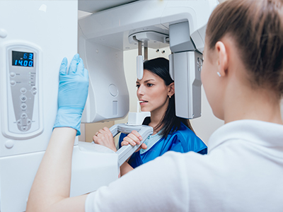
During a digital radiography visit the dental team will position a small sensor inside or near your mouth while you hold still for a few seconds. Most exposures are very quick and cause minimal discomfort thanks to modern sensor design and positioning aids. The number and type of images—bitewings, periapicals, or occlusal views—depend on your oral health needs and the clinical questions your dentist wants to answer.
After capture the image appears almost immediately on a chairside monitor so your clinician can review it with you in real time. If an area needs clarification, software tools can enlarge or adjust the image rather than repeating the exposure. When additional three-dimensional information is required, the dentist may recommend CBCT imaging to supplement two-dimensional radiographs.
Digital radiographs are integral to treatment planning because they provide precise visual information that can be combined with other digital data. Images can be imported into planning software, aligned with intraoral scans, and used alongside three-dimensional CBCT data for implant placement or complex restorative cases. This integration helps clinicians measure space, assess bone quality, and design restorations with predictable outcomes.
Within Restoration Dental these combined digital workflows streamline coordination between the dentist, the dental laboratory, and any specialists involved in care. Shared digital files reduce duplication of imaging and accelerate the move from diagnosis to fabrication of crowns, bridges, or prostheses. As a result, patients experience fewer appointments and a more cohesive treatment timeline.
Digital radiographic records are stored electronically in secure clinical systems that support backups, encryption, and access controls. These safeguards reduce the risk of lost or damaged film and help practices comply with privacy and health record regulations. Authorized staff can retrieve images quickly for follow-ups, emergency visits, or continuity of care when a patient changes providers.
When images need to be transferred for referrals or second opinions, secure electronic methods allow prompt sharing without physical media. Patients can request copies of their radiographs, and many practices provide them in standard formats to support continuity of care. Electronic records also reduce environmental waste by eliminating chemical developers and disposable film.
Yes, digital radiographs are routinely shared with specialists and other providers to support coordinated care. Files can be exported in common image formats and transmitted securely so consultants can review the same information used by your dentist. This accessibility often shortens the time to diagnosis and reduces the need for duplicate imaging.
Before sharing, practices typically obtain patient consent and follow secure transfer protocols to protect privacy. Collaborative review of images facilitates multidisciplinary treatment planning for cases such as implants, oral surgery, or orthodontics. If you are referred, ask your dentist how they will transfer your images and whether they will accompany the referral with a clinical summary.
The frequency of dental x-rays is individualized and based on a patient’s oral health, risk factors, and clinical findings rather than a fixed schedule. New patients often receive baseline images to document current conditions, while recall intervals depend on caries risk, periodontal status, and treatment history. Children and patients with active disease typically require more frequent monitoring than low-risk adults.
Dentists follow professional guidelines and use clinical judgment to balance diagnostic benefit against radiation exposure. The goal is to obtain images only when they will influence diagnosis or treatment, following the principle of keeping exposure as low as reasonably achievable. If you have questions about why a particular image is recommended, your provider can explain the clinical reasoning and alternative options.
Digital radiography offers particular advantages for pediatric patients because sensors and protocols can be adapted to reduce dose and improve comfort. Smaller sensors and faster exposures make the process quicker and less intimidating for children. Lower required radiation and a decreased need for retakes further reduce cumulative exposure during growth and development.
Pediatric imaging focuses on growth, eruption patterns, and early detection of decay to support timely, conservative treatment. Digital records make it straightforward to compare images over time as a child’s dentition changes. Clinicians trained in pediatric care will tailor both the imaging technique and the frequency to each child’s needs.
At Restoration Dental digital radiography is a foundational diagnostic tool that supports rapid, evidence-based decisions at the chairside. Images are integrated with other digital records—such as intraoral scans and CBCT—so the team can plan implants, restorations, and surgical procedures with greater precision. Immediate visualization allows dentists to explain findings clearly and involve patients in treatment decisions.
Staff training, secure record management, and established imaging protocols ensure that radiography contributes to safe and efficient care. By combining lower radiation exposure, image enhancement tools, and streamlined digital workflows, the practice aims to reduce appointment time while improving diagnostic confidence. If you would like more information about how radiographs are used in your care, the dental team can review your images and answer questions at your next visit.
Ready to schedule your next appointment or learn more about our services?
Getting in touch with Restoration Dental is simple! Our welcoming staff is here to help you schedule appointments, answer questions about treatments, and address any concerns you may have. Whether you’d like to call or use our easy online contact form, we’re always ready to assist. Don’t wait to take the first step toward a healthier, more confident smile – contact us today and experience the difference of personalized dental care.